
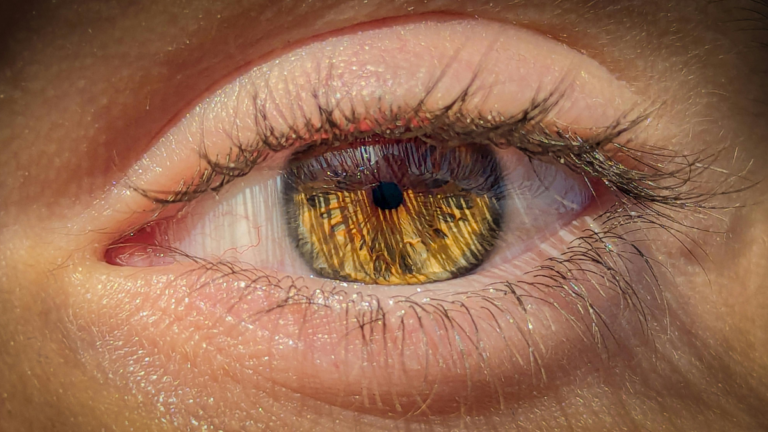

The face finally settled. Six months of consistent treatment, the redness down, the bumps gone, the photographs at last showing something worth keeping. The eyes did not get the message. They still burn by mid-afternoon, still feel like there is sand under the lid, still water in the wind and sting under supermarket lighting. The dermatologist says the rosacea is well controlled. The optician says the eyes look a little dry.
Both are right, and that is the problem. The face and the eye are two separate territories of the same disease, they are graded separately, and improvement in one is not evidence of anything at all in the other.
Quick answer
This is involvement of the eyelids and ocular surface by the same inflammatory disease that affects the facial skin. Reported rates vary widely, with ocular involvement described in up to 72 percent of people with rosacea, and in about 20 percent the eye signs arrive before any skin signs. No reliable correlation has been established between how severe the facial disease looks and how severe the ocular disease is, which means mild skin does not rule out significant eye involvement. The signs that matter sit on the lid margin and need a slit lamp, not a description of symptoms.
The single most useful thing to know
Severity in the skin and severity in the eye are uncoupled. A person with barely visible facial changes can have corneal involvement. A person with florid facial disease can have entirely comfortable eyes. Reviews of the condition state plainly that a correlation between the severity of cutaneous and ocular findings has not been established.

This has one immediate practical consequence. Nobody can reassure you about your eyes by looking at your face. If you have a rosacea diagnosis and any persistent ocular symptoms, the eyes need their own examination, with their own findings recorded and their own treatment plan.
And in one in five people, the eyes come first
The assumption that you need a red face before you can have ocular rosacea is one of the most costly errors in this whole area. Ocular manifestations precede the skin changes in roughly 20 percent of patients, and some series put it nearer 15 percent. Those people spend years being treated for allergic conjunctivitis, chronic dry eye or recurrent styes, because the diagnostic label that would have organised all of it has not arrived yet.
Children are the extreme version of this. In paediatric ocular rosacea the eye signs usually come well before any skin change, delay to diagnosis is more than a year in the large majority of cases, and the consequences include visual impairment and amblyopia. Our guide to Demodex in children covers why any positive finding in a young patient carries more weight than the same finding in an adult.
What the signs actually are
Patients describe symptoms. Symptoms are not specific. Burning, grittiness, foreign body sensation, watering, light sensitivity and morning crusting describe most of ocular surface disease. What identifies this condition is the pattern of physical signs, working from the lid margin inward.

The commonest findings are lid margin telangiectasia, irregular or thickened lid margins and meibomian gland dysfunction, which together drive the evaporative dry eye that dominates day-to-day symptoms. Recurrent chalazia and styes belong on this list too and are frequently the presenting complaint.
The corneal end of the spectrum is what makes this more than a comfort problem. Punctate epithelial erosions have been reported in up to 40 percent of people with the ocular form, and corneal involvement of some degree in up to a third. In a referral series of patients with the ocular form, thirteen presented with reduced vision from corneal complications, six went on to require corneal grafting, and one eye was lost to perforation and infection. That series is a referral population and therefore skewed toward severity, but it establishes the ceiling: this condition has genuine sight-threatening potential when it is left to run.
Where the mite comes into it
The eyelash follicle and the meibomian gland are both sebaceous structures, which makes the lid margin a natural habitat rather than an unusual one. Elevated eyelid margin mite density is a characteristic finding in ocular rosacea, and the associated mechanism has been worked out in more detail here than almost anywhere else in the field.
Mites carry bacteria whose proteins provoke an immune response, and the immunological link has been tested directly. In a study of rosacea patients, ocular mite infestation correlated with serum reactivity to those bacterial proteins. Laboratory work then took the next step and showed that the same proteins induce an abnormal wound-healing response in corneal epithelial cells, which offers a plausible route from a lid margin organism to a corneal ulcer.
Two honest qualifications belong here. First, the classic bacterial candidate in this literature has been reassessed in recent years, and the organism now identified as the Demodex endobacterium is a Corynebacterium species rather than the one named in the older papers, so the mechanism is better described as mite-associated bacterial protein exposure than as a settled single culprit. Second, association is not causation, and a proportion of people with clear ocular rosacea have unremarkable mite counts. The mite is one input, and an addressable one, which is a different claim from being the cause.
Density does appear to track disease extent. In a large retrospective series, facial mite densities measured by consecutive standardised skin surface biopsies were higher in patients who had ocular or scalp involvement, which the authors read as a marker of more advanced disease. A prospective study of papulopustular rosacea found ocular involvement in 40 percent of patients, and reported that follicular scales were significantly more frequent in the mite-positive group.
The gap between two specialties

Dermatology examines the face and asks about the eyes. Ophthalmology examines the eyes and rarely assesses the face. Each is doing its job. The condition spans both.
The size of that gap has been measured. In a cohort of patients with confirmed ocular demodicosis who were also assessed by a dermatologist, 62 percent had concurrent mite overgrowth on the facial skin, and 26 percent had clinically significant facial demodicosis that would have gone undiagnosed and untreated had only the eye examination been performed. In that same cohort, use of topical ivermectin was associated with better resolution of the secondary rosacea features.
If you are the patient standing in that gap, the useful move is to carry the information across it yourself. Tell the eye clinic you have a rosacea diagnosis. Tell the skin clinic your eyes burn. Neither will assume it.
How it is managed
This is a prescription-led condition and the specifics belong with your clinician, but the shape of the plan is worth understanding.
- Lid hygiene is the foundation, and contact time is what makes it work. A five second wipe achieves very little. The lid margin needs sustained contact, and this is the step people abandon first because it looks trivial.
- Oral tetracyclines are the mainstay in moderate to severe disease. They work by damping the inflammatory and protease response rather than by reducing mite numbers, which our guide to doxycycline for rosacea covers in detail, including why the eyes relapse when the tablets stop.
- Mite-directed treatment is a separate lever. Nothing in an oral anti-inflammatory lowers the population on the lid margin.
- Warm compresses and gland expression address the plumbing. They help the blocked oil and do nothing to what is living in the follicle.
- Topical steroid drops are a short-term tool with a long-term cost. They calm inflammation and are sometimes necessary, but local immune suppression is exactly the condition under which mite populations expand, as covered in our article on steroid-induced Demodex.
- Artificial tears treat the symptom, not the disease. Useful for comfort, irrelevant to the lid margin.
Expect the timeline to be measured in months. The follicle cycle described in our life cycle guide is the reason short courses underperform, and the relapse pattern described in our maintenance guide applies to lids exactly as it does to cheeks.
When to stop managing this at home
Most ocular rosacea is a comfort and quality of life problem. A minority is not. Same-week ophthalmology assessment is warranted for any change in vision, significant light sensitivity, eye pain as opposed to irritation, a red eye that is not settling, or a white or hazy patch visible on the cornea. Contact lens wearers with new persistent redness should be assessed promptly rather than assuming lens intolerance.
Anyone whose eye symptoms have run for months through repeated treatment for allergy or dry eye without a lid margin examination should ask directly whether ocular rosacea and mite density have been considered. Our self-assessment is a reasonable place to organise what you have noticed before that appointment, and the practitioner directory lists clinicians who work with this condition.
For practitioners
Three points change management. First, decouple the grading. The absence of an established correlation between cutaneous and ocular severity means facial improvement cannot serve as a proxy endpoint for the ocular surface, and both need independent assessment at each review. The 2019 global consensus defines cutaneous and ocular features separately and recommends combination therapy where multiple features coexist.
Second, note the classification constraint that produces diagnostic delay. Under the phenotype criteria, ocular manifestations are explicitly not independently diagnostic of rosacea, unlike persistent centrofacial erythema or phymatous change. A patient presenting with isolated ocular disease therefore fails the criteria on ocular findings alone, which is a structural reason for the observed lag rather than simple oversight. The consensus does note that ocular rosacea can be identified even with minimal skin involvement through lid margin abnormalities, corneal findings or scleral inflammation.
Third, examine the face. In the ocular demodicosis cohort described above, 62 percent had concurrent facial overgrowth and 26 percent had clinically significant facial demodicosis detectable only on dermatological assessment. A brief facial inspection for follicular scaling, background erythema and textural change adds little time and changes the treatment plan in a meaningful fraction of cases. Additional material is collected in our practitioner resources.
Frequently asked questions
Can you have eye involvement without facial rosacea?
Yes. Ocular involvement precedes any skin change in about 20 percent of patients, and the ocular form can be diagnosed in the presence of minimal skin findings. This is a major reason the condition is missed, since many clinicians look for a red face first.
Does treating my facial rosacea fix my eyes?
Not reliably. No correlation has been established between the severity of the facial findings and the severity of the ocular findings, and oral anti-inflammatory treatment acts on the inflammatory response rather than on the lid margin population. The eyes usually need their own targeted plan.
Can ocular rosacea damage your sight?
It can, though most cases do not. Corneal involvement of some degree has been reported in up to a third of patients, and referral series include people who lost vision to corneal complications. Prompt assessment of any vision change, eye pain or non-settling redness is the safeguard.
Is it Demodex or is it rosacea?
In practice this is often a false choice. Eyelid margin mite density is characteristically raised in this condition, and mite-associated bacterial proteins have been shown to trigger an abnormal wound-healing response in corneal epithelial cells. The mite is best understood as one addressable input into the inflammation rather than as an alternative diagnosis.
Why do artificial tears not help much?
Because they replace the tear film without changing what is disrupting it. The underlying problem is at the lid margin, in the meibomian glands and the lash follicles, so drops improve comfort for an hour or two while the source continues unchanged.
How long does treatment take to work?
Longer than most people are told. Meaningful change is measured in months rather than weeks, partly because the follicle population turns over on roughly a two-week cycle and partly because lid margin inflammation settles slowly. Stopping at the first improvement is the commonest reason it returns.
References
- Geng RSQ, Bourkas AN, Mufti A, Sibbald RG. Ocular rosacea: the often-overlooked component of rosacea. JEADV Clinical Practice. 2024;3(3):723-732.
- Tan J, Almeida LMC, Bewley A, et al. Updating the diagnosis, classification and assessment of rosacea: recommendations from the global ROSacea COnsensus (ROSCO) panel. British Journal of Dermatology. 2017;176(2):431-438. PubMed
- Schaller M, Almeida LMC, Bewley A, et al. Recommendations for rosacea diagnosis, classification and management: update from the global ROSacea COnsensus 2019 panel. British Journal of Dermatology. 2020;182(5):1269-1276. PubMed
- Akpek EK, Merchant A, Pinar V, Foster CS. Ocular rosacea: patient characteristics and follow-up. Ophthalmology. 1997;104(11):1863-1867.
- Fromstein SR, Harthan JS, Patel J, Opitz DL. Demodex blepharitis: clinical perspectives. Clinical Optometry. 2018;10:57-63.
- Li J, O’Reilly N, Sheha H, et al. Correlation between ocular Demodex infestation and serum immunoreactivity to Bacillus proteins in patients with facial rosacea. Ophthalmology. 2010;117(5):870-877.
- O’Reilly N, Gallagher C, Reddy Katikireddy K, et al. Demodex-associated Bacillus proteins induce an aberrant wound healing response in a corneal epithelial cell line: possible implications for corneal ulcer formation in ocular rosacea. Investigative Ophthalmology and Visual Science. 2012;53(6):3250-3259.
- Forton FMN, De Maertelaer V. Rosacea and demodicosis: little-known diagnostic signs and symptoms. Acta Dermato-Venereologica. 2019;99(1):47-52.
- Trave I, Cozzani E, Salvi I, Parodi A. Follicular scales, scalp and ocular involvement in patients with papulopustular rosacea: prevalence and association with Demodex mite proliferation. Dermatology Reports. 2023;15(3).
- Chirinos-Saldana P, Bonifaz A, et al. Comorbidity of ocular and facial demodicosis. American Journal of Ophthalmology. 2023;255:100-108.
- Oltz M, Check J. Rosacea and its ocular manifestations. Optometry. 2011;82(2):92-103.
About the author. Marina Ivakhnenko is a functional skin practitioner who works with people whose rosacea, blepharitis and folliculitis have not responded to standard care. This article is educational and is not a substitute for individual medical advice. Any change in vision, eye pain or a red eye that is not settling needs assessment by an eye clinician, not a website.


