
Introduction
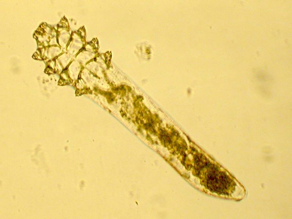
Demodex mites are microscopic parasites that naturally inhabit human hair follicles and sebaceous glands. While small populations are usually harmless, excessive Demodex proliferation can contribute to various skin and eye conditions, including rosacea, blepharitis, folliculitis, and persistent facial irritation.
Because Demodex mites are invisible to the naked eye, accurate diagnosis requires specialized examination techniques. Among the available diagnostic methods, Demodex Dermoscopy has emerged as a valuable, non-invasive tool for identifying signs of Demodex infestation.
This article explores how Dermoscopy, specifically Demodex Dermoscopy, is used in Demodex diagnosis, what clinicians look for, and how it compares to other testing methods.
What Is Dermoscopy?
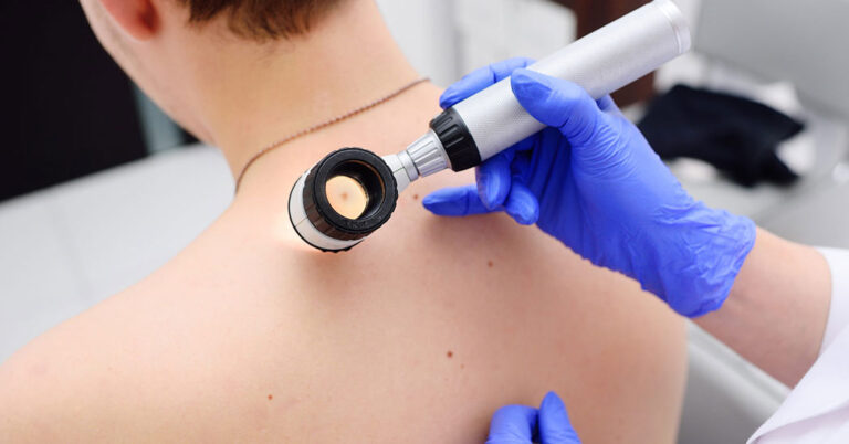
Dermoscopy is a non-invasive imaging technique that uses a handheld device called a dermatoscope to magnify and illuminate the skin.
The technique allows dermatologists to examine structures beneath the skin surface that cannot be seen with standard visual inspection.
Dermoscopy is commonly used for:
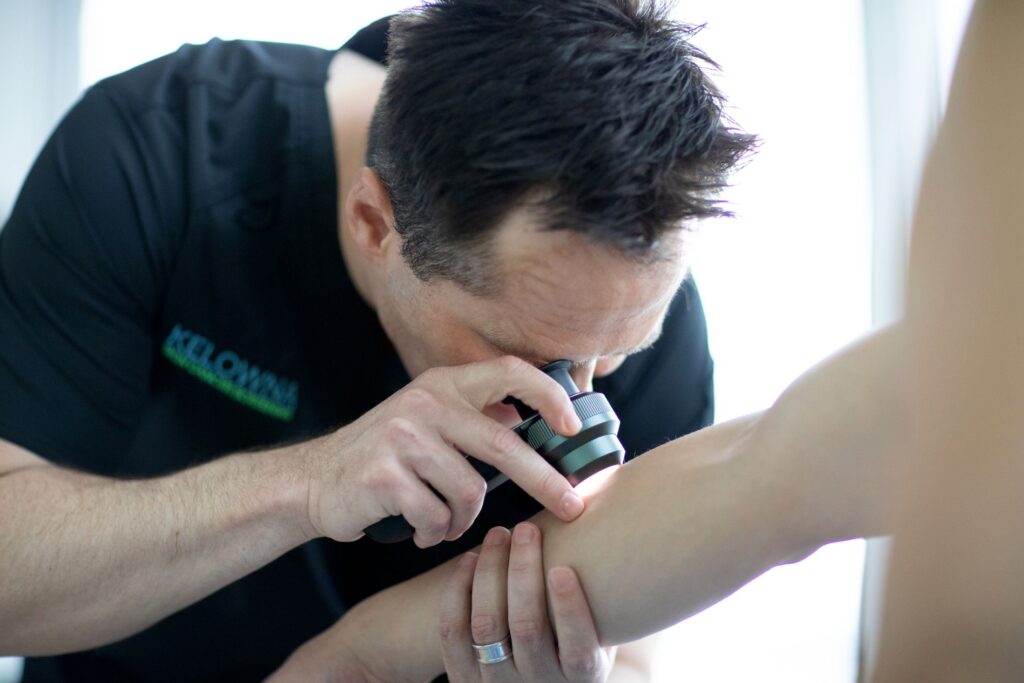
Because it does not require skin scraping or biopsy, dermoscopy is often the first diagnostic step when Demodex infestation is suspected.
Signs of Demodex Seen Under Dermoscopy
Dermoscopy cannot directly identify every individual mite, but it reveals characteristic features associated with high Demodex density.
1. Demodex Tails
One of the most distinctive dermoscopic findings is the presence of “Demodex tails.”
These appear as:
Demodex tails represent accumulated keratin and mite waste protruding from infested follicles.
2. Follicular Openings
Affected follicles often appear:
Clusters of enlarged follicles may indicate significant mite colonization.
3. Follicular Scaling
Fine scales surrounding hair follicles are another common dermoscopic sign.
These scales may indicate:
4. Vascular Changes
Patients with rosacea-related Demodex overgrowth may demonstrate:
These findings help clinicians distinguish Demodex-associated inflammation from other skin conditions.
Conditions Where Dermoscopy Is Helpful
Rosacea
Research has shown that patients with rosacea often have higher densities of Demodex mites than healthy individuals.
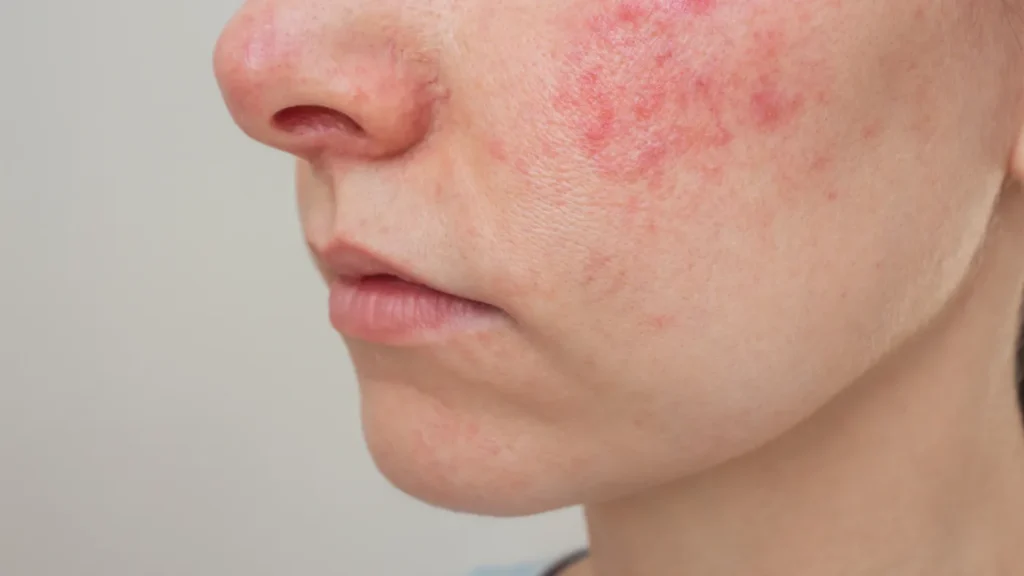
Dermoscopy may help identify:
Blepharitis
For eyelid Demodex infestation, clinicians may observe:
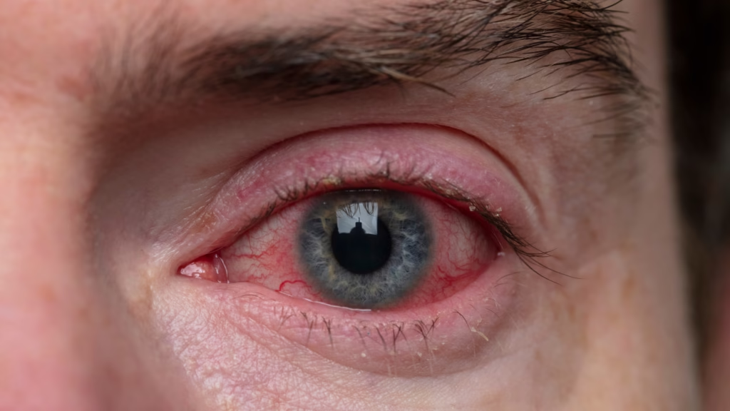
These findings frequently correlate with chronic blepharitis.
Folliculitis
Persistent folliculitis that does not respond to conventional treatment may be associated with Demodex overgrowth.
Dermoscopy can reveal characteristic follicular changes suggesting mite involvement.
Other Diagnostic Methods for Demodex
While dermoscopy is highly useful, it is often combined with additional testing methods.
Standardized Skin Surface Biopsy (SSSB)
This technique involves applying adhesive to a microscope slide and sampling the skin surface.
Advantages:
Limitations:
Skin Scraping
A clinician gently scrapes the affected area and examines the sample under a microscope.
Advantages:
Limitations:
Eyelash Sampling
For suspected ocular Demodex infestation, several eyelashes may be removed and examined microscopically.
This method is commonly used in cases of chronic blepharitis and recurrent chalazia.
Confocal Laser Scanning Microscopy
This advanced imaging technology allows real-time visualization of mites within hair follicles.
Advantages:
Limitations:
Benefits of Dermoscopy for Demodex Diagnosis
Dermoscopy offers several advantages:
These benefits make dermoscopy an increasingly popular first-line diagnostic tool in dermatology clinics.
Limitations of Dermoscopy
Although dermoscopy is valuable, it does have limitations.
It cannot:
For definitive diagnosis, dermatologists may combine dermoscopy with microscopic testing.
Monitoring Treatment Success
Dermoscopy is also useful during treatment.
Follow-up examinations can help clinicians assess:
This allows more objective evaluation of treatment effectiveness.
Conclusion
Dermoscopy has become an important tool for diagnosing Demodex infestations. By revealing characteristic features such as Demodex tails, enlarged follicles, and follicular scaling, it helps clinicians identify mite-related skin disorders quickly and non-invasively.
Although microscopic methods remain the gold standard for confirming Demodex density, dermoscopy provides an excellent first-line assessment and can play a key role in monitoring treatment progress. For patients with persistent rosacea, blepharitis, or unexplained follicular inflammation, dermoscopic evaluation may offer valuable insights into the possible role of Demodex mites.
References
- Usefulness of Dermoscopy in the Diagnosis and Monitoring Treatment of Demodicidosis
- Dermoscopy as a Diagnostic Tool in Demodicidosis
- Dermoscopic Findings of Rosacea and Demodicosis
- Demodex Mite Density Determinations by Standardized Skin Surface Biopsy and Direct Microscopic Examination
- Demodex Blepharitis: A Comprehensive Review of the Disease, Diagnosis, and Treatment
- Demodex, demodicosis — DermNet NZ


