
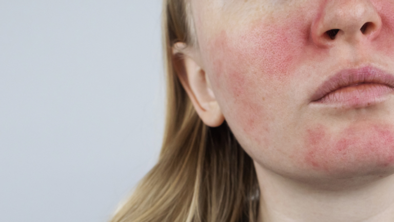

Almost everyone arrives at this question the same way. Someone said the word rosacea, you went looking, and within two clicks you were reading about four numbered types and trying to work out which one you are. Then the answer would not settle. You have the redness from type one and the bumps from type two. Or the nose changes from type three with none of the pustules. Or gritty eyes and a face that looks broadly fine.
You are not failing to understand the system. The system was retired, by the same expert bodies that created it, because it did exactly this to real patients.
Quick answer
The four rosacea types came from a 2002 classification: type 1 erythematotelangiectatic, type 2 papulopustular, type 3 phymatous, type 4 ocular. In 2017 both the global ROSacea COnsensus panel and the National Rosacea Society moved to a phenotype approach, because individual features routinely span more than one subtype and the boxes were confounding both research and treatment. Under the current criteria two features are diagnostic on their own: persistent centrofacial erythema that periodically intensifies, and phymatous change. Flushing, telangiectasia, papules and pustules, and ocular manifestations are not individually diagnostic. The practical shift is that you are treated feature by feature, not by category.
What the four rosacea types were
In 2002 a National Rosacea Society expert committee published the first standard classification, sorting the common patterns of signs into four groupings.
- Subtype 1, erythematotelangiectatic. Persistent central facial redness with flushing and visible blood vessels.
- Subtype 2, papulopustular. Redness with the inflammatory bumps and pustules that get mistaken for acne.
- Subtype 3, phymatous. Thickening of the tissue, most often the nose, with enlarged pores and irregular contour.
- Subtype 4, ocular. Involvement of the eyelids and ocular surface.
The system was genuinely useful. It gave a shared vocabulary, made trials comparable, and pulled a poorly defined condition into focus. Its authors were explicit that it was provisional and expected to need revision as the science matured.
It is worth being clear about what happened next, because the language online has not caught up. The rosacea types were not disproved and they were not abandoned in confusion. They were superseded, deliberately, by the same kind of expert committee that produced them, once fifteen years of clinical use had exposed where they broke down. Older articles, older leaflets and a great deal of current search content still teach the numbers as though nothing changed.
Why it stopped working

The central problem was overlap. Individual features span multiple rosacea types, so the same person legitimately belongs in more than one box at once. Centrofacial redness, for instance, appears in the definition of both subtype 1 and subtype 2, which means epidemiology could never be sure whether a patient with redness and papules had been counted once or twice. Severity scoring inherited the same confusion, because grading a subtype means grading several different features at the same time and hoping they move together. They do not.
There was also a narrative built into the numbering that the evidence never supported. The sequence implies progression, from redness through bumps to thickening, and patients read it as a prognosis. Reviews of the classification debate note there has not been convincing evidence that people progress from one end of the spectrum to the other. Having redness now does not mean a thickened nose later. That single correction relieves more anxiety than almost anything else on this page.
The mite evidence points the same way

A meta-analysis pooling 23 case-control studies and 1,513 patients found significantly higher mite density in rosacea than in controls, with an odds ratio of 9.039 for infestation. When the analysis was broken down by subtype, the standardised mean difference in density was 2.686 for the erythematotelangiectatic presentation and 2.804 for the papulopustular one. Those are very close numbers for two categories that were supposed to be meaningfully different.
That does not prove the subtypes were meaningless, and mite density is only one variable among several. But it fits the direction the classification moved in: the visible differences between presentations reflect how a given person’s skin responds, more than they reflect a different disease process underneath.
How rosacea is diagnosed now

The 2017 global consensus, reached by seventeen dermatologists and three ophthalmologists using a blinded Delphi process, established that two features are independently diagnostic:
- Persistent centrofacial erythema associated with periodic intensification. Redness across the central face that is always there to some degree and gets worse in episodes.
- Phymatous changes. Thickened tissue, most often on the nose.
Flushing, telangiectasia, inflammatory lesions and ocular manifestations were specifically judged not to be individually diagnostic. Any two of them in combination will support the diagnosis, but none alone settles it. The National Rosacea Society update published in the same period took a compatible line, retiring the subtypes as units for diagnosis and treatment while allowing the old groupings to be referred to descriptively as neurovascular, inflammatory, phymatous and ocular.
This is where a lot of diagnostic delay comes from. Someone whose only problem is burning, gritty eyes does not meet the criteria on ocular findings alone, which is one structural reason isolated eye involvement waits so long for a name. That situation is covered in our guide to ocular rosacea.
What gets mistaken for rosacea
Because the diagnosis is clinical and the features are shared with other conditions, the differential matters more than the numbering ever did.
- Acne vulgaris. The discriminator is comedones. Blackheads and closed comedones belong to acne and are not a feature of rosacea. Acne also tends to include lesions at several different stages at once.
- Seborrhoeic dermatitis. Greasy scale in the nasolabial folds, eyebrows and hairline, with flaking rather than papules. The two coexist often enough that finding one does not exclude the other.
- Perioral dermatitis. Clustered small papules around the mouth with a characteristic clear rim at the lip border, very often following topical steroid use.
- Demodicosis. The mite-driven presentation overlaps so heavily with the papulopustular picture that some authors consider them the same disease. Practical pointers toward mite involvement include fine follicular scaling, a rough or sandpaper texture, diffuse background redness between the bumps, and burning or stinging out of proportion to how the skin looks. Our article on rough skin texture covers the driest version of this, which is also the most frequently missed.
What this changes for you
Dropping the numbers is not a technicality. It changes what a good consultation looks like.
- The right question is which features you have, not which of the rosacea types you are. Persistent redness, flushing episodes, visible vessels, papules and pustules, thickening, eye involvement, plus the burning and stinging that often go unrecorded. Write your own list before the appointment.
- Each feature has its own treatment. Anti-inflammatory agents address papules and pustules. Vasoconstrictors address background redness. Light-based treatment addresses visible vessels. None of these substitutes for the others, which is why one prescription rarely handles a mixed picture.
- Combination therapy is expected, not a sign of failure. The 2019 update explicitly recommends combination treatment for people with multiple features.
- The goal is complete clearance, not almost clear. The 2019 panel set complete clearance as the primary treatment objective, on the basis of demonstrated benefit to patients. If your treatment stalls at improved but not clear, that is a reason to keep going rather than to accept it.
- What drives the features still needs asking about. Feature-led treatment is a better map than the subtypes, but it is still a description of what shows. The question of what is provoking it sits underneath, and mite density is one of the few parts of that answer that can actually be measured. Our self-assessment is a starting point.
Where treatment has controlled the visible features but relapse keeps arriving on a timetable, our guides to maintenance and doxycycline explain why suppression and resolution are different things. For a broader view of the evidence base, see our research section.
For practitioners
The transition is now well documented and worth stating plainly to patients who arrive with a subtype label from the internet or from an older record. ROSCO 2017 achieved consensus for phenotype-based diagnosis and classification, identifying persistent centrofacial erythema with periodic intensification and phymatous change as independently diagnostic, with flushing, telangiectasia, inflammatory lesions and ocular manifestations explicitly not individually diagnostic. The NRS 2017 update aligned, reframing the former subtypes as descriptive groupings linked to pathophysiology rather than as diagnostic units.
Three consequences for practice. First, severity assessment should be per feature rather than per category, since subtype grading conflates features that respond to different agents and change at different rates. Second, the 2019 update sets complete clearance rather than near clearance as the primary objective and recommends combination therapy where multiple features coexist, which reframes partial response as an indication to escalate rather than an acceptable endpoint. Third, the progression narrative embedded in the numbering is not supported by convincing evidence, and correcting it is a meaningful intervention in its own right for patients who believe phymatous change is inevitable.
On the mite question, the Chang and Huang meta-analysis reported an odds ratio of 9.039 for infestation across 23 case-control studies with near-identical standardised mean differences in density between the erythematotelangiectatic and papulopustular groups, at 2.686 and 2.804 respectively. Whatever weight is given to Demodex causally, the density data do not respect the subtype boundary. Further material is in our practitioner resources.
Frequently asked questions
What are the four rosacea types?
The four rosacea types come from a 2002 classification, which described subtype 1 erythematotelangiectatic, meaning redness and visible vessels; subtype 2 papulopustular, meaning bumps and pustules; subtype 3 phymatous, meaning thickened tissue; and subtype 4 ocular, meaning eye involvement. Expert panels moved away from this system in 2017 in favour of diagnosing and treating by individual feature.
Which of the rosacea types do I have?
Under current criteria that is no longer the question clinicians are asked to answer. Features regularly span more than one of the old categories, which is precisely why the system was replaced. What matters is the list of features you actually have, because each one is treated differently.
Does rosacea progress from type 1 to type 3?
The numbering implies a sequence, but there has not been convincing evidence that patients progress from one end of the spectrum to the other. Having persistent redness now does not mean phymatous change is coming later.
Can you have more than one type of rosacea at once?
Yes, and that overlap is the reason the rosacea types were retired as diagnostic categories. A person can have persistent redness, inflammatory lesions and eye involvement simultaneously, which made assigning a single category arbitrary and confounded both severity scoring and research.
How is rosacea diagnosed if not by type?
By phenotype. Either persistent centrofacial erythema with periodic intensification or phymatous change is diagnostic on its own. Flushing, telangiectasia, papules and pustules, and ocular manifestations each support the diagnosis but need at least one other feature alongside them.
Is Demodex a separate condition from rosacea?
The relationship is close enough that some authors treat mite-driven disease and the papulopustular presentation as the same entity. Pooled analysis found a substantially higher likelihood of infestation in rosacea than in controls, with similar density elevations across the redness-dominant and bump-dominant presentations. Whether it is called a driver or a co-traveller, it is one of the few parts of the picture that can be measured directly.
References
- Tan J, Almeida LMC, Bewley A, et al. Updating the diagnosis, classification and assessment of rosacea: recommendations from the global ROSacea COnsensus (ROSCO) panel. British Journal of Dermatology. 2017;176(2):431-438. PubMed
- Gallo RL, Granstein RD, Kang S, et al. Standard classification and pathophysiology of rosacea: the 2017 update by the National Rosacea Society Expert Committee. Journal of the American Academy of Dermatology. 2018;78(1):148-155. PubMed
- Schaller M, Almeida LMC, Bewley A, et al. Recommendations for rosacea diagnosis, classification and management: update from the global ROSacea COnsensus 2019 panel. British Journal of Dermatology. 2020;182(5):1269-1276. PubMed
- Wilkin J, Dahl M, Detmar M, et al. Standard classification of rosacea: report of the National Rosacea Society Expert Committee on the Classification and Staging of Rosacea. Journal of the American Academy of Dermatology. 2002;46(4):584-587.
- Schaller M, Almeida LMC, Bewley A, et al. Rosacea treatment update: recommendations from the global ROSacea COnsensus (ROSCO) panel. British Journal of Dermatology. 2017;176(2):465-471.
- Chang YS, Huang YC. Role of Demodex mite infestation in rosacea: a systematic review and meta-analysis. Journal of the American Academy of Dermatology. 2017;77(3):441-447.
- Tan J, Berg M. Rosacea: current state of epidemiology. Journal of the American Academy of Dermatology. 2013;69(6 Suppl 1):S27-S35.
- Forton FMN. Papulopustular rosacea, skin immunity and Demodex: pityriasis folliculorum as a missing link. Journal of the European Academy of Dermatology and Venereology. 2012;26(1):19-28.
- Tan J, Blume-Peytavi U, Ortonne JP, et al. An observational cross-sectional survey of rosacea: clinical associations and progression between subtypes. British Journal of Dermatology. 2013;169(3):555-562.
- Gether L, Overgaard LK, Egeberg A, Thyssen JP. Incidence and prevalence of rosacea: a systematic review and meta-analysis. British Journal of Dermatology. 2018;179(2):282-289.
About the author. Marina Ivakhnenko is a functional skin practitioner who works with people whose rosacea, folliculitis and blepharitis have not responded to standard care. This article is educational and is not a substitute for individual medical advice. Diagnosis of rosacea is clinical and belongs with a clinician who can examine your skin.


