You have tried three cleansers, two prescription creams, and a handful of supplements. The redness keeps coming back. The morning grittiness in your eyes is now a daily event. Something itches on your face at night that nothing else explains. You searched the obvious diagnoses, and now you are looking at a far less obvious one: microscopic mites that may have been living in your facial follicles for years.
This guide answers the practical question patients arrive at when standard treatments stop working. How do you tell if you have Demodex mites? The answer is not a single test. It is a pattern of signs, a structured self-assessment, and a professional confirmation step that takes minutes in a properly equipped clinic.
What You Are Actually Looking For
Demodex are microscopic mites that live inside the hair follicles and sebaceous glands of human skin. They are part of normal skin biology. Most adults carry small numbers without ever knowing it.
The condition is not the presence of the mite. The condition is overgrowth, when mite density rises above the level the local immune system can quietly manage. This threshold sits at around five mites per square centimetre of facial skin on objective testing. Above that threshold, the immune response shifts into a chronic inflammatory pattern that produces the visible signs patients look up online.
Two Demodex species affect humans. Demodex folliculorum measures 0.3 to 0.4 mm and lives in hair follicle openings, especially around the eyelashes, nose, and cheeks. Demodex brevis is smaller at 0.15 to 0.2 mm and lives deeper inside sebaceous and meibomian glands. The two species drive subtly different patterns. D. folliculorum dominates the visible facial signs. D. brevis dominates the deeper eyelid and gland problems that produce dry eye and texture changes.

The Two-Layer Framework for Recognising Demodex Overgrowth
Telling if you have Demodex involves two layers of evidence that need to point in the same direction.
Layer 1: Symptom pattern. Demodex overgrowth has a recognisable clinical signature. It produces a specific set of symptoms in a specific anatomic distribution with a specific timing pattern. No single symptom is diagnostic on its own. A combination of several, especially the seven hallmark signs covered below, raises the clinical suspicion significantly.
Layer 2: Treatment-resistance pattern. The strongest single predictor of underlying Demodex is incomplete response to well-executed standard therapy for whatever surface diagnosis you have already received. Patients who have been treated for rosacea, blepharitis, or perioral dermatitis with appropriate first-line therapy and who keep relapsing are the highest-yield Demodex testing candidates in dermatology and ophthalmology clinics.
The five hallmark signs, the seventeen-point self-assessment, and the five professional diagnostic tests reviewed below operate across both layers. Most patients with confirmed demodicosis show evidence in at least three categories.
The Five Hallmark Signs of Demodex Overgrowth
01. Persistent Central Facial Redness That Does Not Respond Fully to Standard Rosacea Therapy
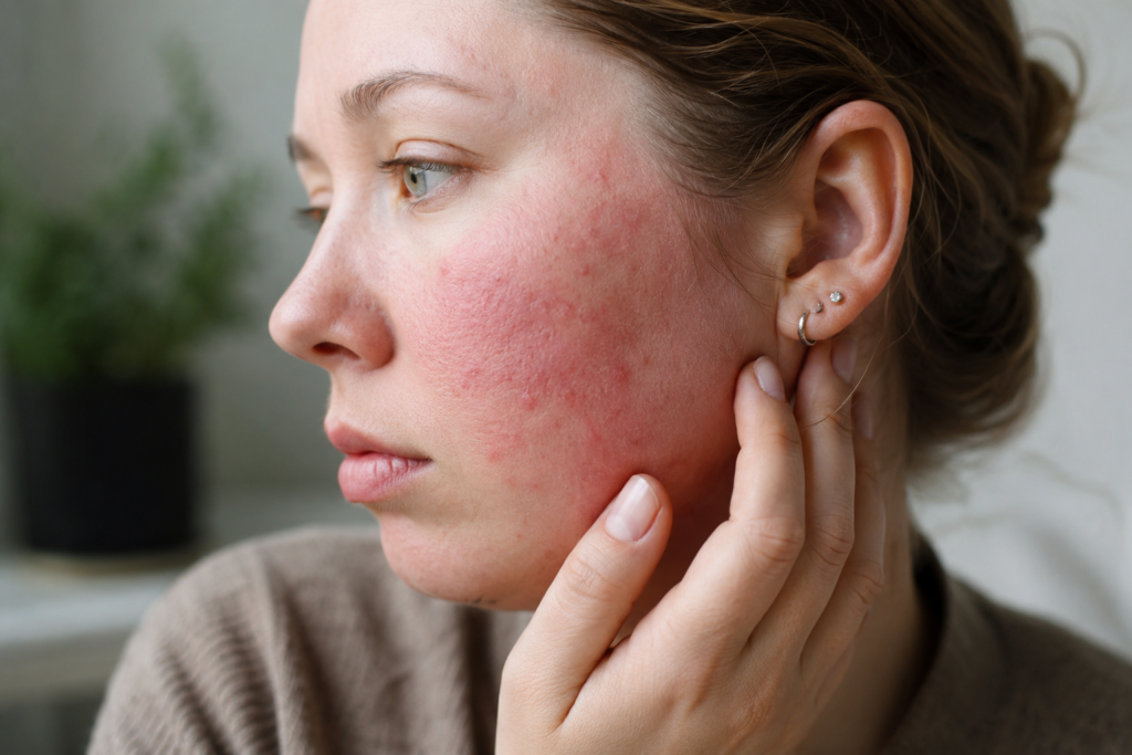
What you may notice. Sustained pink or red colouring across the cheeks, nose, and central forehead that has been present for months or years. The redness may be background and constant, or it may flare in episodes triggered by alcohol, hot drinks, exercise, or stress.
Why it points to Demodex. The mite triggers inflammation in the central facial pilosebaceous units by activating toll-like receptor 2 (TLR2) on keratinocytes. The downstream cathelicidin pathway then produces the chronic vasodilation and redness that characterises this presentation. Demodex-associated rosacea is now considered a major contributor to papulopustular and erythematotelangiectatic rosacea subtypes.
The diagnostic clue. If you have been using topical metronidazole, azelaic acid, or oral doxycycline for rosacea, and the redness either does not clear completely or returns within weeks of stopping, the Demodex layer is unlikely to have been targeted.
02. Sandpaper Skin Texture and Visible Follicular Plugging
What you may notice. Skin that feels rough to the touch despite a moisturising routine. Enlarged or conspicuous pores, particularly on the nose and cheeks. Small comedo-like plugs in the nasal alar creases.
Why it points to Demodex. Mite density inside the follicle drives reactive hyperkeratinisation, which is what produces the gritty texture. The mite waste and shed exoskeletons solidify at the follicle opening, producing the visible plugging.
The diagnostic clue. The texture often persists even when the surface inflammation looks calmer. Exfoliating acids reduce it temporarily but do not resolve it because they do not address the cause. The texture returns within days of stopping the exfoliant.

03. Eyelid Itching, Grittiness, and Waxy Buildup at the Lash Base
What you may notice. Eyelids that feel itchy, burning, or gritty, especially on waking. Recurrent styes or chalazion. Eyelashes that look thinner than they used to be. Crusty, waxy collars wrapped around the base of the eyelashes, visible on close inspection in good light.
Why it points to Demodex. The cylindrical waxy collars (clinically called collarettes or cylindrical dandruff) are made of solidified mite waste, eggs, and shed exoskeletons. They are considered pathognomonic for Demodex blepharitis. Their presence on slit lamp examination is essentially diagnostic.
The diagnostic clue. If your eyelid symptoms have been treated as ordinary dry eye or allergic conjunctivitis without improvement, the next step is a slit lamp examination by an eye care provider who knows to look for collarettes. Diagnosis takes under two minutes once the patient looks downward.
04. Symptoms That Worsen at Night
What you may notice. Itching, burning, or a crawling sensation on the face or scalp that intensifies after going to bed. Eyelid symptoms that feel worst on waking. Sleep that is interrupted by the urge to rub or scratch the face.
Why it points to Demodex. Demodex mites are photophobic. They emerge from the follicular openings onto the skin surface at night to mate. The increase in surface mite activity drives a measurable increase in local inflammatory mediators during the hours of sleep. This nocturnal pattern is one of the most distinctive symptom features and is rarely seen in primary rosacea or allergic skin disease.
The diagnostic clue. If your skin or eyelid symptoms have a consistent daily rhythm that is worst at night and on waking, Demodex deserves serious consideration regardless of the surface diagnosis you have been given.
05. Symptoms Triggered or Worsened by Topical Corticosteroids
What you may notice. A clear pattern in which skin or eyelid symptoms emerged shortly after starting a topical steroid (for any reason), or in which symptoms became worse during a course of facial steroid use, or in which symptoms rebound aggressively after stopping a steroid.
Why it points to Demodex. Topical corticosteroids suppress the local skin immune response that normally restrains Demodex density. Sustained steroid use on the face can cause mite density to rise dramatically. The resulting condition (steroid-induced rosacea or perioral dermatitis) often has a substantial Demodex component that goes unrecognised because the inflammation is attributed solely to the steroid effect.
The diagnostic clue. This pattern is common in patients who used topical steroids long term for eczema, seborrhoeic dermatitis, or non-specific facial irritation. If your symptoms have a clear temporal relationship with topical steroid exposure, Demodex testing is appropriate.
The Seventeen-Point Self-Assessment Checklist
Mark every statement that applies to you. The combination of items, not any individual item, is what matters. Bring the completed list to your dermatologist or ophthalmologist appointment.
Skin symptoms
- I have persistent central facial redness or flushing.
- My skin texture feels rough, uneven, or sandpaper-like.
- I have recurring pustules, papules, or follicular bumps.
- My pores look enlarged, particularly on the nose and cheeks.
- My skin reacts badly to products that used to be fine.
- I have visible follicular plugging or comedo-like lesions.
- My skin symptoms started or worsened after topical steroid use.
Eye and eyelid symptoms
- My eyelids itch, burn, or feel gritty.
- I have noticed waxy collars at the base of my eyelashes.
- I have had recurrent styes, chalazion, or eyelid bumps.
- My eyes feel dry, watery, or sensitive to light.
- I have noticed eyelash thinning or eyelash loss.
Timing and pattern
- My symptoms feel worse at night or on waking.
- I have a sensation of crawling or movement on my skin.
- I have a diagnosis of rosacea, blepharitis, or perioral dermatitis that has not fully responded to first-line treatment.
- My symptoms have lasted more than three months despite treatment.
- I have a condition or treatment that suppresses my immune system, or I am over forty years old.
The Five Professional Tests That Confirm Demodex

No home test reliably confirms Demodex overgrowth. Diagnosis is established in a clinical setting using one or more of the following methods.
Test 1: Standardised skin surface biopsy (SSSB). A drop of cyanoacrylate glue is applied to the skin, a glass slide is pressed onto it, and the slide is gently peeled away once the glue has set. The slide is then examined under a microscope. Mite count is reported per square centimetre. A density above five per square centimetre is considered elevated. This is the most widely used quantitative dermatology test for Demodex.
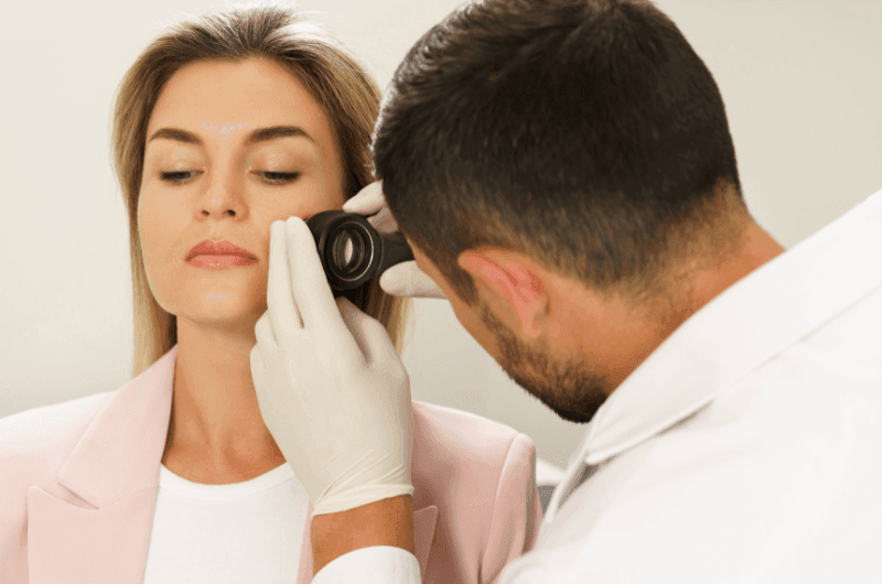
Test 2: Dermoscopy. A handheld or videodermoscope at high magnification reveals mite tails protruding from follicular openings, follicular plugging, and the so-called rosette or “Demodex tail” pattern. This is the quickest in-office screening method and is the standard initial assessment in most contemporary dermatology clinics.
Test 3: Eyelash epilation with light microscopy. An ophthalmologist or optometrist removes several lashes (typically three to four from each eyelid) and examines them under direct light microscopy. More than one to two mites per lash is considered diagnostically significant for Demodex blepharitis. This remains the standard method in ophthalmology.
Test 4: Reflectance confocal microscopy (RCM). A non-invasive in vivo imaging technique that visualises mites within the follicle in real time. RCM has the highest sensitivity of any clinical method but is restricted to specialist dermatology centres that have the equipment.
Test 5: Slit lamp examination for collarettes. The patient looks downward at a slit lamp while the clinician evaluates the upper lid margin. Cylindrical waxy collars at the base of the lashes are considered essentially diagnostic for Demodex blepharitis. The Cataract and Refractive Surgery Today consensus reports that 100 percent of patients with visible collarettes have Demodex blepharitis. The examination takes under two minutes.
Why Some People Are More Susceptible Than Others
Most adults carry low-density Demodex without symptoms. Overgrowth happens when something tips the balance. Identifying which of the following applies to you helps your clinician understand the picture and helps you reduce the chance of recurrence after treatment.
- Age over forty. Mite density increases progressively with age. Most adults over fifty carry meaningfully more Demodex than they did in their twenties, even without symptoms.
- Immune suppression. Any cause of reduced cellular immunity, including HIV, leukaemia, organ transplantation, chemotherapy, or systemic immunosuppressant medication, allows mite density to rise.
- Topical corticosteroid use on the face. Even short courses of mid-potency facial steroids can produce noticeable Demodex blooms in susceptible patients.
- Diabetes mellitus. Associated with elevated mite density and more refractory presentations. Glycaemic control matters.
- High sebum production. The mite is sebum-dependent. Oily skin types and patients with hormonal drivers of sebum production carry higher baseline densities.
- Compromised skin barrier. Eczematous skin, over-exfoliated skin, and skin with chronic low-grade inflammation all show elevated Demodex counts.
- Gut microbiome disturbance. The Demodex-gut axis is increasingly recognised as a systemic driver of recurrent demodicosis. Small intestinal bacterial overgrowth (SIBO) prevalence in rosacea patients exceeds forty percent.
- Alcohol consumption. Identified as the single strongest modifiable lifestyle factor in 2024 cohort data, with an odds ratio above eleven for Demodex-associated dermatosis.
Further Reading on demodex.net/
- Demodex Blepharitis: The Complete Clinical Guide to Eyelid Mite Disease
- Demodex and Rosacea: The Complete Clinical Guide to the Mite-Rosacea Connection
- Tea Tree Oil for Demodex Mites: A Complete Clinical Evidence Guide
- Natural Remedies for Demodex Mites: A Clinical Evidence Review
- The Demodex-Gut Axis: Why Your Skin Mites Are Really a Gut Health Problem
- Demodex Research Library: Peer-Reviewed Evidence
- Find a Demodex-Aware Practitioner Near You
Frequently Asked Questions
Can I see Demodex mites with the naked eye?
No. Adult Demodex folliculorum measures around 0.3 millimetres and Demodex brevis is even smaller at 0.15 to 0.2 millimetres. Both are well below the limit of unaided human vision, which is approximately 0.1 millimetres for high-contrast objects under ideal lighting and considerably more for low-contrast translucent organisms inside skin follicles. What you can see without magnification are the consequences of Demodex overgrowth, not the mites themselves: collarettes at the lash base, follicular plugging, papules, and persistent erythema. Direct visualisation of the mite requires dermoscopy, light microscopy, or reflectance confocal microscopy.
Is there a reliable at-home test for Demodex mites?
No validated at-home test exists at the time of writing. Online resources occasionally describe taping methods or smartphone microscopy attempts, but these do not produce clinically interpretable results and are not recommended. The closest you can come at home is structured symptom assessment using the seventeen-point checklist above, photographic documentation of any visible signs (particularly collarettes on the upper eyelashes in good lighting), and tracking the timing pattern of your symptoms. These observations make a professional examination substantially more productive.
How long does it take to get a Demodex diagnosis confirmed?
In most cases, under five minutes during a clinic visit, once you are with a clinician who knows to look. Dermoscopy and slit lamp examination for collarettes are immediate. A standardised skin surface biopsy takes about ten minutes total including microscope examination. Eyelash epilation with microscopy takes approximately the same. The bottleneck is usually finding a clinician who routinely considers Demodex in the differential, which is why our practitioner directory exists.
Will my regular doctor know how to test for Demodex?
Probably not at primary care level. Demodex assessment is generally a dermatology or ophthalmology procedure. Most primary care physicians are aware of the condition but do not have the equipment (dermoscope, slit lamp, microscope) needed for direct confirmation. If you suspect Demodex, the most efficient route is to ask your primary care provider for a referral to a dermatologist for skin-predominant symptoms or to an ophthalmologist or optometrist for eyelid-predominant symptoms.
Can children have Demodex mites?
Children carry far lower Demodex densities than adults, and symptomatic demodicosis is uncommon before puberty. When facial or eyelid demodicosis is identified in a child or adolescent, it warrants investigation for an underlying cause of immune compromise. Otherwise unexplained paediatric demodicosis is a recognised red flag and should not be managed as a routine skin condition without further assessment.
If I have one Demodex-related condition, am I likely to have others?
Yes, more often than not. The risk factors that allow overgrowth at one body site usually allow overgrowth at others. Patients with confirmed Demodex blepharitis frequently also have facial Demodex involvement, and patients with Demodex-associated rosacea frequently have low-grade Demodex blepharitis that has not yet been examined for. A complete assessment looks at the face, the eyelids, and the scalp together, regardless of which symptom originally prompted the appointment.
Are Demodex mites contagious between people?
Mite transfer between close contacts (partners, parents and children, anyone sharing pillowcases or eye makeup) is documented in clinical research. The transfer alone does not produce disease. Most recipients carry the transferred mites at low density without symptoms. Disease develops only when individual host factors (immune regulation, skin barrier, sebaceous activity, systemic inflammation, age) allow mite overgrowth. So you cannot give someone rosacea or blepharitis by skin contact, even though you can transfer mites.
What should I do if my dermatologist dismisses Demodex as a possibility?
Two reasonable next steps. First, bring the completed seventeen-point checklist and ask specifically for a dermoscopic examination of your central face and for an eyelash microscopy or slit lamp examination if eyelid symptoms are present. These tests are quick and inexpensive. Second, if your dermatologist will not perform or refer for these tests, seek a second opinion from a clinician with documented experience in Demodex. The demodex.net/ practitioner directory lists providers with this experience by region.
References and Evidence Sources
- Forton FMN, De Maertelaer V. Two consecutive standardized skin surface biopsies: an improved sampling method to evaluate Demodex density as a diagnostic tool for rosacea and demodicosis. Acta Dermato-Venereologica. 2017;97:242-248.
- Ayres BD, Donnenfeld E, Farid M, et al. Clinical diagnosis and management of Demodex blepharitis: the Demodex Expert Panel on Treatment and Eyelid Health (DEPTH). Eye. 2023;37:3249-3255.
- Chang YS, Huang YC. Role of Demodex mite infestation in rosacea: a systematic review and meta-analysis. Journal of the American Academy of Dermatology. 2017;77(3):441-447.e6.
- Trattler W, Karpecki P, Rapoport Y, et al. The prevalence of Demodex blepharitis in US eye care clinic patients as determined by collarettes: a pathognomonic sign. Clinical Ophthalmology. 2022;16:1153-1164.
- Lacey N, Russell-Hallinan A, Powell FC. Study of Demodex mites: challenges and solutions. Journal of the European Academy of Dermatology and Venereology. 2024;38(2):245-255.
- Sędzikowska A, Osęka M, Grytner-Zięcina B. Ocular symptoms reported by patients infested with Demodex mites. Acta Parasitologica. 2016;61(4):808-814.
- Friedman P, Sabban EC, Cabo H. Usefulness of dermoscopy in the diagnosis and monitoring of demodicosis. Dermatology Practical and Conceptual. 2017;7(1):35-38.
- Forton FMN. Papulopustular rosacea, skin immunity and Demodex: pityriasis folliculorum as a missing link. Journal of the European Academy of Dermatology and Venereology. 2012;26(1):19-28.
- Demirseren DD, Cicek H, Aksoy Sarac G. Which factors influence Demodex mite density in standardized superficial skin biopsy in patients with rosacea? A prospective cross-sectional analysis. PubMed PMID 38787449. 2024.
- Geng RSQ, Bourkas AN, Mufti A, Sibbald RG. Rosacea: pathogenesis and therapeutic correlates. Journal of Cutaneous Medicine and Surgery. 2024;28(2):178-189.
- Rather PA, Hassan I. Human Demodex mite: the versatile mite of dermatological importance. Indian Journal of Dermatology. 2014;59(1):60-66.
- Parodi A, Paolino S, Greco A, et al. Small intestinal bacterial overgrowth in rosacea: clinical effectiveness of its eradication. Clinical Gastroenterology and Hepatology. 2008;6(7):759-764.
- Tabriz University of Medical Sciences Cohort. A cross-sectional survey of the relationship between rosacea and Demodex mite infestation. Iranian Journal of Dermatology. 2025;28(1):e123456.
- EOS Study Investigators. Proportion of patients with Demodex blepharitis in ophthalmology clinics in Europe: the EOS study. Eye. 2025;39:adv4130.
- Wolffsohn JS, Arita R, Chalmers R, et al. TFOS DEWS II Diagnostic Methodology report. The Ocular Surface. 2017;15(3):539-574.
About the Author
Marina Ivakhnenko is a clinical writer and editorial lead at demodex.net/, where she develops evidence-based content for patients and practitioners managing Demodex mite overgrowth. Her work draws from peer-reviewed dermatology, ophthalmology, and parasitology literature and is reviewed by the demodex.net/ clinical advisory board. Marina specialises in translating complex parasitology and skin immunology research into clear, clinically actionable guidance for both patient and practitioner audiences.